GHK-Cu is a copper-binding tripeptide with significant preclinical evidence in wound healing and skin regeneration. In laboratory settings, it activates fibroblasts to increase collagen and elastin production, modulates matrix metalloproteinases for balanced tissue remodeling, and promotes angiogenesis. It also suppresses inflammatory markers such as IL-6 and TNF-alpha while influencing the expression of more than 4,000 genes tied to repair. Definitive human efficacy data is not yet available, and the sections below explain why that distinction matters.
What Is GHK-Cu and Why Does It Matter for Skin?
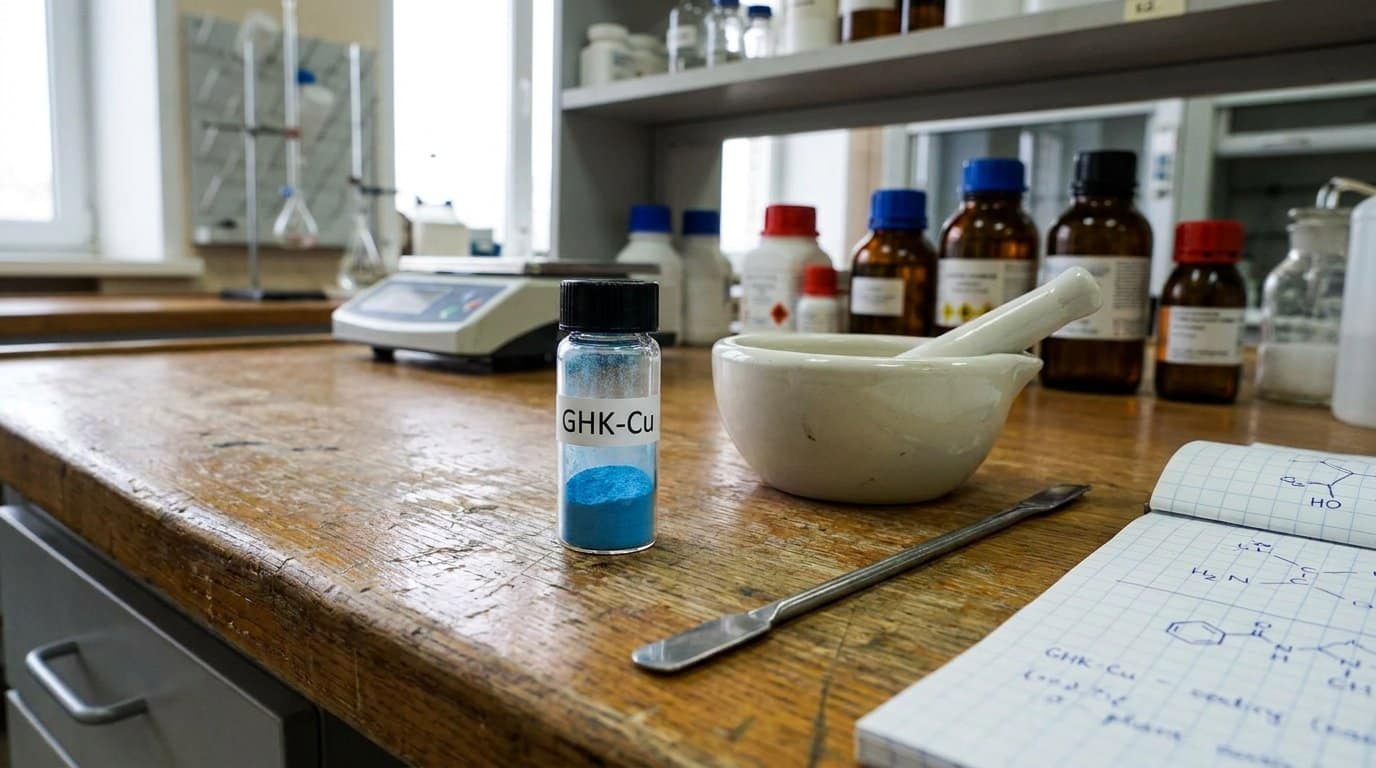
GHK-Cu, glycine-histidine-lysine bound to copper(II) and also designated copper tripeptide-1, is a naturally occurring signaling molecule found in human plasma, saliva, and urine. Copper binding amplifies its biological activity, making the complex form functionally distinct from the unbound peptide. GHK-Cu is not a synthetic-only construct. It is part of native biochemistry, operating as a broad tissue-maintenance signal.
In skin biology, GHK-Cu skin research findings point to measurable effects on fibroblasts, keratinocytes, and dermal remodeling pathways. Laboratory evidence links GHK-Cu to increased collagen synthesis, glycosaminoglycan production, and gene-expression shifts across thousands of targets. Review literature consistently describes it as a regenerative signaling molecule rather than a surface-level cosmetic agent, with documented activity in inflammation modulation, oxidative stress response, and cellular repair programming. Laboratory studies have reported that GHK-Cu can increase collagen production by up to 70% under controlled conditions, reinforcing its significance as a measurable driver of dermal matrix remodeling rather than a passive ingredient.
How GHK-Cu Repairs Skin: Collagen, Blood Vessels, and Cell Signaling
GHK-Cu fibroblast research shows upregulated synthesis of collagen, elastin, and glycosaminoglycans, which strengthens dermal architecture in study models. At the same time, the peptide modulates MMPs and their inhibitors, balancing matrix degradation against rebuilding.
| Mechanism | Key Outcome |
|---|---|
| Fibroblast activation | Increased collagen, elastin, decorin production |
| MMP modulation | Balanced extracellular matrix remodeling |
| Angiogenesis promotion | Enhanced blood vessel formation in injured tissue |
| Gene expression shifts | Modulation of >4,000 genes, including IL-6 and TNF-alpha suppression |
Angiogenic effects do not operate in isolation. They are coupled with connective tissue rebuilding, creating coordinated vascular and structural repair that accelerates wound closure in models. The peptide also acts as an anti-inflammatory agent by lowering pro-inflammatory cytokines, supporting an environment that favors tissue regeneration.
GHK-Cu and Wound Healing: What Animal Studies Show
Before GHK-Cu‘s cellular mechanisms were mapped in detail, animal wound models had already established its capacity to accelerate tissue repair across multiple species and injury types. Rabbit studies showed faster wound contraction, increased angiogenesis, and elevated antioxidant enzyme activity. Rodent models extended these findings, showing systemic repair effects beyond the local application site.
GHK-Cu wound healing research has also documented effects in impaired-healing contexts. Ischemic rat wounds with restricted blood supply and diabetic wound models both showed accelerated closure, reduced inflammatory burden, and enhanced collagen synthesis. GHK-Cu also improved skin graft take rates and supported pad wound healing in dogs. Across open, ischemic, diabetic, and graft models, preclinical data consistently indicate improved closure, contraction, and tissue remodeling quality. These findings align with work by Pickart and Margolina exploring the peptide’s regenerative and protective actions in connection with gene expression and potential applications.
Antioxidant and Anti-Inflammatory Effects of GHK-Cu
Beyond its direct effects on wound closure and tissue remodeling, GHK-Cu shows significant antioxidant and anti-inflammatory activity relevant to its wound-healing mechanisms. Laboratory studies show GHK-Cu inactivates lipid peroxidation by-products, including 4-hydroxynonenal, acrolein, and malondialdehyde, while protecting cultured keratinocytes from UV-induced oxidative damage through neutralization of these reactive aldehydes. In parallel, GHK-Cu suppresses NF-κB-mediated inflammatory signaling and reduces TNF-α levels across multiple injury models, indicating that the peptide’s tissue-protective profile extends well beyond structural protein synthesis.
Lipid Peroxidation Inactivation
Because oxidative stress generates not only primary reactive oxygen species but also secondary reactive aldehydes, including 4-hydroxynonenal, acrolein, malondialdehyde, and glyoxal, cellular damage in wounded or inflamed tissue extends well beyond the initial oxidative insult. These aldehydes modify proteins, disrupt membranes, and impair signaling pathways critical to repair. Acrolein, for example, inactivates Cu,Zn-superoxide dismutase through histidine loss and copper release, compounding redox imbalance.
GHK-Cu directly binds and inactivates these reactive by-products before they reach cellular targets. In wound model research, this aldehyde-sequestering activity protects fibroblasts and keratinocytes from secondary oxidative injury, preserving the cellular machinery required for collagen synthesis and tissue remodeling. By reducing reactive aldehyde burden at the site of injury, GHK-Cu limits downstream protein modification and supports more efficient progression through wound repair phases.
UV Radiation Protection
While GHK-Cu‘s aldehyde-sequestering capacity addresses secondary oxidative damage in wound environments, the same mechanism extends to ultraviolet radiation injury in skin keratinocyte models. In vitro studies report that GHK-Cu protects keratinocytes from lethal UVB exposure through direct antioxidant activity and reactive carbonyl species inactivation.
Key findings from ghk-cu skin aging research on UV defense include:
- ROS neutralization, where GHK-Cu blocks reactive oxygen species generated immediately after UVB irradiation
- Inflammatory cascade suppression, where the peptide reduces post-UV inflammatory mediator signaling
- Endogenous defense upregulation, where gene-expression shifts favor antioxidant and repair pathway activation
- Cumulative stress recovery, where cellular defense mechanisms support resilience against chronic environmental damage
It is worth noting that GHK-Cu does not function as a sunscreen. Its UV-related role in these models is adjunctive post-exposure support, not SPF-based photoprotection.
TNF-Alpha Reduction Effects
Among GHK-Cu’s documented anti-inflammatory mechanisms, TNF-α suppression is one of the most consistently reproduced findings across both cell culture and animal models. In LPS-stimulated RAW 264.7 macrophages, GHK-Cu reduced TNF-α production alongside ROS levels while increasing SOD activity and GSH concentrations. Mechanistically, this suppression involved inhibition of NF-κB p65 and p38 MAPK signaling pathways.
In DSS-induced colitis mouse models, GHK-Cu lowered TNF-α and other pro-inflammatory cytokines, including IL-6 and IL-1β, through SIRT1/STAT3 pathway modulation. These TNF-α reduction effects are directly relevant to ghk-cu scar research, since sustained TNF-α elevation drives chronic inflammation that impairs collagen remodeling. By shifting tissue from a pro-inflammatory state toward repair, GHK-Cu’s TNF-α-lowering activity supports the downstream regenerative processes central to wound resolution.
Skin Remodeling, Hair Growth, and Other Emerging Findings
Beyond its direct wound-closure effects, GHK-Cu exerts measurable influence on broader skin remodeling processes, including stimulation of fibroblast-driven collagen deposition, increased glycosaminoglycan synthesis, and enhanced decorin production, all of which contribute to organized extracellular matrix architecture and improved tissue integrity. In parallel, published preclinical data link GHK-Cu to increased hair follicle size and thicker hair shaft diameter, effects likely mediated through the same growth factor signaling and tissue repair pathways that underpin its wound healing activity. While the skin remodeling evidence draws support from both cell culture and in vivo models, the hair growth findings remain largely preclinical and require further controlled investigation to establish reproducibility and relevance.
Skin Remodeling Effects
Although GHK-Cu‘s wound-healing properties have received the most sustained research attention, its skin remodeling effects represent a distinct and increasingly documented area of investigation. GHK-Cu laboratory findings consistently point to broad pathway modulation rather than a single-target mechanism, with dermal fibroblasts serving as a central cellular target.
Key remodeling effects documented in the experimental literature include:
- Increased collagen and elastin synthesis in dermal fibroblast cultures
- Matrix metalloproteinase regulation supporting balanced matrix turnover alongside new formation
- Enhanced glycosaminoglycan production, including dermatan sulfate and chondroitin sulfate
- Gene-expression shifts associated with a healthier repair phenotype across multiple signaling pathways
These effects are driven by GHK-Cu’s copper-binding signaling activity, which coordinates matrix rebuilding and controlled remodeling at the same time.
Hair Growth Research
GHK-Cu’s effects on hair follicle biology represent a more recent but growing branch of its skin research literature. Preclinical models show GHK-Cu accelerates the shift from telogen to anagen phase through activation of the Wnt/β-catenin signaling pathway and upregulation of follicle-related growth factors, including Ldha expression in mouse skin.
Beyond direct follicle stimulation, copper peptide wound healing studies have identified VEGF-mediated microcirculation improvements that enhance nutrient delivery to follicular structures. Researchers have documented increased follicle size, hair thickness, and epithelial cell proliferation in treated specimens. GHK-Cu’s anti-inflammatory and antioxidant properties further support follicle viability by reducing oxidative damage in the perifollicular microenvironment. This evidence remains mainly preclinical, with delivery method optimization representing a critical variable in observed outcomes.
Is GHK-Cu Proven? What Clinical Trials Tell Us So Far
Despite decades of promising preclinical data, human clinical evidence for GHK-Cu‘s wound-healing effects remains limited. Most ghk-cu dermal research derives from cell culture and animal models rather than controlled human trials.
A registered clinical study is now testing topical GHK-Cu gel for acute skin wound healing. The reported design includes:
- Population: healthy adults with small, standardized skin wounds
- Intervention: topical gel containing GHK-Cu versus matching vehicle
- Primary outcome: time to complete re-epithelialization
- Goal: establish safety and measurable healing acceleration
GHK-Cu is biologically active and well-supported preclinically, but definitive human efficacy data do not yet exist. It is best regarded as a promising investigational peptide rather than a proven wound-healing therapy.
Shop Research Peptides at Holas Today
If you are looking for laboratory-grade peptides that are properly handled, securely packaged, and shipped with care, Holas has you covered. We provide research-grade peptides with third-party tested purity, reliable packaging standards, and fast shipping to support your research needs. Browse our full catalog or contact us to find the right materials for your research today. All products are for laboratory research use only and are not for human or veterinary use.
Frequently Asked Questions
How Does GHK-Cu Compare to Other Copper-Binding Peptides in Research?
GHK-Cu has greater research depth and breadth than other copper-binding peptides. It has the most replicated evidence across wound healing, collagen synthesis, elastin support, and anti-inflammatory activity in both cell and animal models. AHK-Cu, by contrast, has a narrower research footprint with fewer cited mechanistic studies. Most conclusions about copper peptides in skin biology are derived primarily from GHK-Cu data, which makes it the established experimental benchmark.
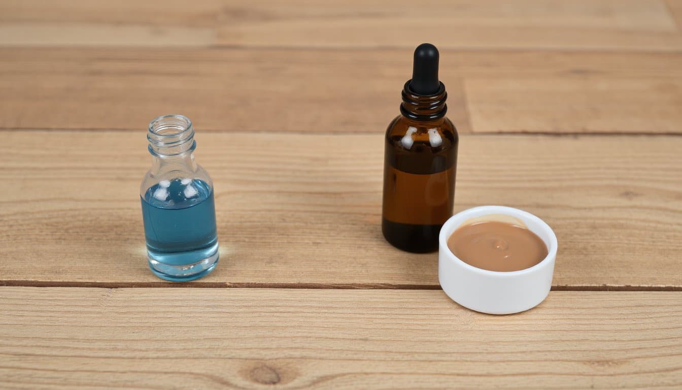
What Delivery Methods Work Best for GHK-Cu in Laboratory Studies?
Hydrogel-based delivery systems have shown the strongest performance in laboratory studies, outperforming simple solution application by prolonging wound-bed contact time and sustaining local bioactivity. Topical gels and polymer-encapsulated formulations consistently improved wound closure rates and biochemical outcomes in both in vitro and in vivo models. Nanoparticle carrier systems and pH-sensitive conjugates further enhanced stability and penetration, while injectable routes appear more relevant to systemic regenerative research than to localized applications.
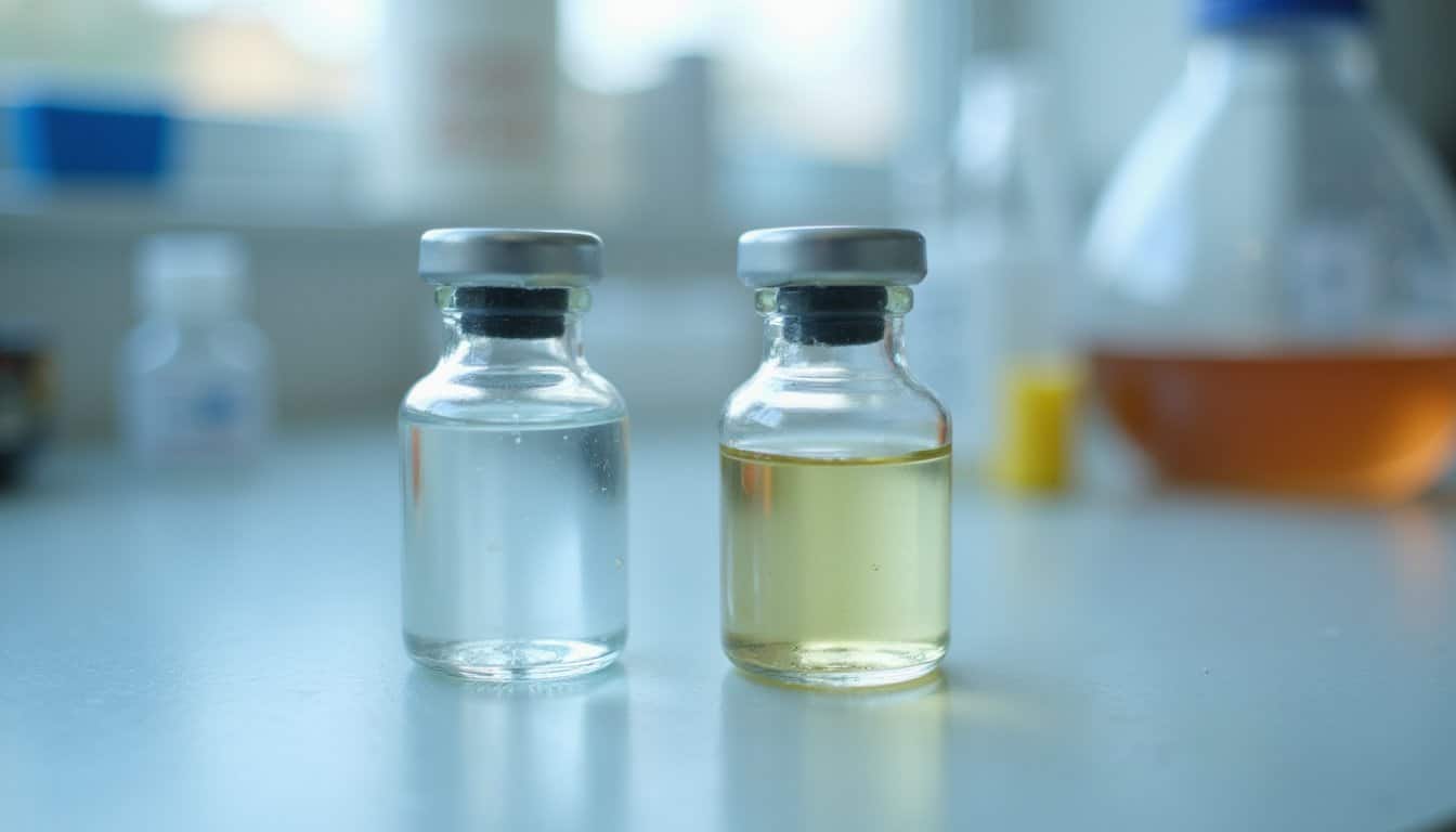
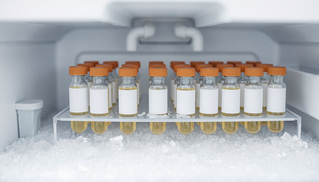
Does GHK-Cu Stability Vary Across Different pH or Temperature Conditions?
Yes. GHK-Cu’s stability varies markedly with both pH and temperature. The copper-peptide complex degrades faster under strongly acidic or alkaline conditions, with most formulation data targeting a pH range of roughly 5.0 to 6.5 for improved stability. Temperature is equally important, as reconstituted solutions degrade faster at room temperature than under refrigeration at 2 to 8°C. Lyophilized powder maintains stability considerably longer, especially when stored frozen and sealed.
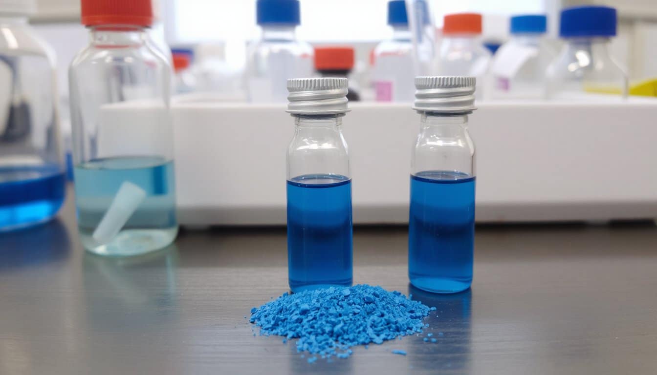
Can GHK-Cu Interact Negatively With Other Compounds in Formulations?
Yes. In formulation research, GHK-Cu can interact unfavorably with specific compounds. Ascorbic acid destabilizes the copper complex through oxidation, reducing both ingredients’ measured activity. Retinoids combined with GHK-Cu have been associated with increased irritation potential in formulation testing. pH mismatches and oxidative interactions in multi-ingredient blends further degrade stability. These issues are typically managed in study design by separating incompatible actives and characterizing each component independently before combining them.
What Concentration of GHK-Cu Is Used in Cell Studies?
The concentration depends on the endpoint. For collagen I stimulation in dermal fibroblasts, 1 nM GHK-Cu has repeatedly shown the strongest response, with collagen production peaking at this low dose and declining at higher concentrations. For keratinocyte stemness markers such as integrins and p63, studies typically use low micromolar concentrations. Dose-response behavior is non-linear, so higher concentrations do not necessarily improve results, and the literature emphasizes that GHK-Cu’s activity peaks at very low doses.