GHK-Cu activates dermal fibroblasts and increases synthesis of type I and type III collagen by up to 70% in laboratory models, partly through TGF-beta signaling, at concentrations as low as 0.01 nM. It also modulates more than 4,000 human genes, upregulating pathways tied to extracellular matrix assembly and suppressing those linked to destructive inflammation. Copper’s role as a lysyl oxidase cofactor further supports collagen cross-linking. The sections below break down each mechanism and the evidence behind it.
What Is GHK-Cu and Why Do Researchers Study It?
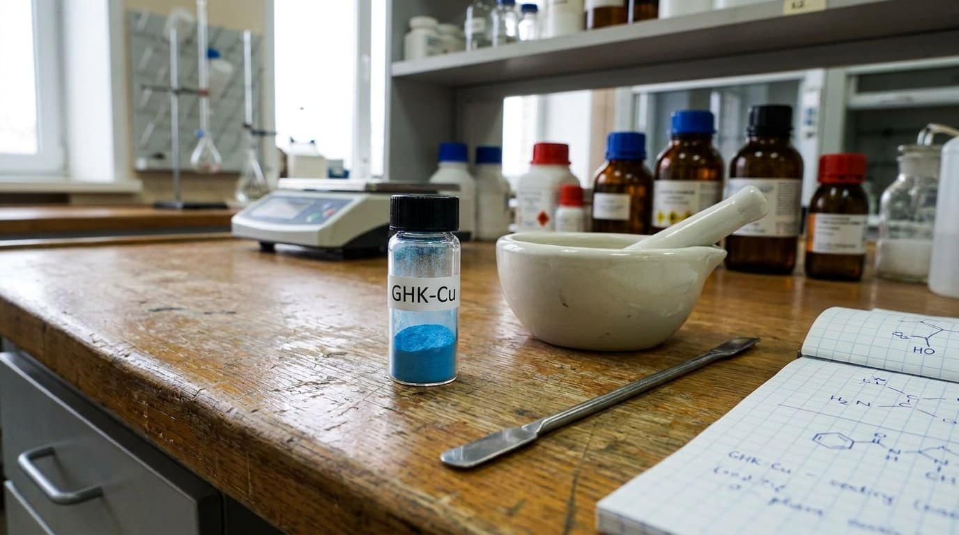
GHK-Cu is a naturally occurring copper(II) complex of the tripeptide glycyl-L-histidyl-L-lysine, first isolated from human plasma in the early 1970s. It is present in saliva, urine, and circulating plasma, where its levels decline measurably with age. This age-related decline, particularly notable after age 60, correlates with the decreased regenerative capacity observed in older adults.
Researchers study GHK-Cu because it functions as a multifaceted signaling molecule across damage response and repair pathways. GHK-Cu collagen research shows its capacity to stimulate synthesis of types I, III, and IV collagen at nanomolar concentrations. Studies examining GHK-Cu gene expression have documented modulation of more than 4,000 genes spanning tissue remodeling, anti-inflammatory, and antioxidant pathways. GHK-Cu fibroblast activation drives extracellular matrix production, including glycosaminoglycans and decorin. These converging mechanisms make it a recurring target in wound healing, regenerative medicine, and dermatological research.
How GHK-Cu Triggers Collagen Production in Skin Cells
When human dermal fibroblasts are exposed to GHK-Cu, the peptide activates these cells to upregulate collagen synthesis through TGF-beta signaling pathway stimulation, with measurable effects at concentrations as low as 0.01 nM. Cultures show increased production of both collagen types I and III, the primary structural and reparative collagens in skin, alongside enhanced fibroblast proliferation that amplifies overall matrix output. This dual mechanism, combining direct collagen gene upregulation with expansion of the collagen-producing cell population, establishes the foundation for the extracellular matrix remodeling effects measured in downstream tissue assays. Laboratory studies have reported that GHK-Cu can increase collagen production by up to 70% under controlled conditions, underscoring the potency of this peptide’s effect on dermal matrix biosynthesis.
Fibroblast Activation Mechanisms
GHK-Cu also activates fibroblasts through EGFR transactivation, promoting migration and differentiation signaling that supports organized tissue repair. Acting as a chemoattractant, GHK-Cu stimulates fibroblast proliferation and migration toward wound sites, supporting sufficient cellular density for extracellular matrix production. Beyond receptor-level events, gene regulation and epigenetic remodeling underlie GHK-Cu’s broader fibroblast reprogramming, shifting aged or damaged cells toward functional repair phenotypes. This transcriptional breadth distinguishes GHK-Cu from single-pathway growth factors in wound healing models.
Collagen Synthesis Pathways
At concentrations as low as 1 to 10 nM, the copper-bound tripeptide drives a selective increase in collagen secretion from human dermal fibroblasts without proportionally elevating total non-collagen protein output. This selectivity distinguishes ghk-cu collagen production from generalized protein upregulation, pointing to pathway-specific activation rather than broad translational stimulation.
Copper peptide collagen production depends partly on copper’s role as a lysyl oxidase cofactor, directly supporting collagen cross-linking and maturation. GHK-Cu also upregulates MMP1, MMP2, and TIMP1 expression, coordinating synthesis with controlled remodeling. Decorin induction further organizes nascent collagen fibrils into functional architecture. The ghk-cu 4000 genes research confirms these are not isolated effects. Collagen pathway genes operate within a broader transcriptomic shift favoring matrix assembly, balanced turnover, and structured tissue repair.
GHK-Cu’s Effects on Type I and Type III Collagen
GHK-Cu’s collagen-stimulating activity differs between type I and type III collagen, which serve structurally distinct roles in dermal tissue. Type I provides tensile strength in mature skin, while type III predominates in early-phase wound repair and vascular structures. Cell culture studies in human adult dermal fibroblasts confirm that GHK-Cu directly increases collagen production at concentrations as low as 0.01 nM, with gene expression data showing coordinated upregulation of extracellular matrix assembly pathways that encompass both collagen subtypes alongside glycosaminoglycans and decorin. Type I collagen has been measured as a direct endpoint in multiple fibroblast and biopsy-based studies, while type III collagen support is inferred primarily through GHK-Cu’s activation of broader connective tissue remodeling and early wound-healing signaling pathways rather than isolated quantification in most available datasets.
Collagen Type Differences
Because collagen is not a single molecule but a family of structurally distinct proteins, the specific types GHK-Cu upregulates determine its functional impact on tissue architecture. Type I collagen provides tensile strength and long-term dermal firmness, while type III collagen dominates early granulation tissue during repair. GHK-Cu’s influence on both types reflects its dual role in acute healing and structural maintenance.
The ghk-cu wound healing mechanism involves coordinated type III deposition during initial repair, followed by type I replacement during maturation. This shift depends on ghk-cu tgf-beta signaling, which regulates fibroblast differentiation and matrix protein selection. In parallel, ghk-cu elastin research shows the peptide does not act on collagen in isolation. It co-stimulates elastin production, so the remodeled matrix retains both rigidity and elastic recoil rather than favoring one mechanical property.
Fibroblast Synthesis Evidence
| Parameter | Observation |
|---|---|
| Active concentration range | 10⁻¹² to 10⁻⁹ M |
| Peak response | ~10⁻⁹ M |
| Collagen types increased | Type I and Type III |
| Proliferation dependence | None detected |
| Remodeling character | Organized, not fibrotic |
Rat wound models corroborate these in vitro findings, showing elevated collagen I and III expression following GHK-Cu injection, which reinforces translational relevance beyond cell culture systems.
How GHK-Cu Shifts Gene Activity Toward Repair and Renewal
Although GHK-Cu’s collagen-stimulating activity is its most studied function, the peptide’s influence on gene expression extends far beyond any single protein target. Large-scale profiling studies have identified more than 4,000 human genes modulated by GHK-Cu, revealing a consistent pattern: upregulation of repair-associated gene sets and downregulation of damage- and inflammation-associated expression.
The data show increased transcriptional activity in genes governing extracellular matrix maintenance, antioxidant defense, cellular migration, and angiogenesis signaling. At the same time, GHK-Cu suppresses gene sets linked to destructive inflammation, fibrosis, and chronic stress responses. This dual regulatory action does not target a single receptor-ligand axis but coordinates multiple pathways at once. Review articles characterize the shift as a partial transcriptional reset toward healthier expression profiles in damaged or aged cells, favoring resolution-oriented repair over prolonged inflammatory activation.
How GHK-Cu Rebuilds the Skin’s Support Structure
The gene expression shifts described above set the stage for GHK-Cu‘s most functionally visible outcome: physical reconstruction of the dermal extracellular matrix. In fibroblast models, GHK-Cu increases type I collagen and elastin synthesis at nanomolar concentrations, strengthening the dermal scaffold through active remodeling rather than transient volumization.
GHK-Cu also modulates matrix metalloproteinases and upregulates tissue inhibitors of metalloproteinases, favoring net matrix repair over degradation. Studies show increased glycosaminoglycan output, including dermatan sulfate, chondroitin sulfate, and decorin, which stabilizes collagen fiber organization and restores the ground substance these fibers depend on.
At the basement membrane level, GHK-Cu supports collagen IV production, reinforcing the epidermal-dermal junction. Combined with enhanced angiogenesis and fibroblast proliferation, these mechanisms produce measurable wound closure acceleration and connective tissue reconstruction in preclinical models.
What Human Skin Studies Reveal About GHK-Cu and Collagen
While preclinical fibroblast and wound models establish GHK-Cu’s collagen-stimulating mechanisms at the cellular level, human skin studies provide the translational data on whether these effects produce measurable structural changes in living tissue. In a 12-week facial cream trial involving 71 women with photoaging, researchers documented increased skin density and thickness, reduced laxity, and fewer fine lines. A separate IRB-approved trial in 21 women reported that subdermal echogenic density, a validated proxy for collagen and elastin content, rose by an average of 28% over three months of daily topical application, with top-quartile responders reaching 51%.
Comparative analyses add further detail. A 2022 randomized controlled trial reported a wrinkle reduction of roughly 55.7% with 1% GHK-Cu cream versus about 32.2% for the vehicle control, with histological confirmation of increased dermal collagen. Many human studies remain small-scale and short-term, often summarized within review literature rather than large independent trials.
How GHK-Cu Compares to Vitamin C and Retinoids
Because GHK-Cu, vitamin C, and retinoids all influence collagen metabolism through distinct mechanisms, comparing them requires examining each compound’s primary mode of action rather than treating them as interchangeable. In a human thigh-skin biopsy study, GHK-Cu increased collagen production in 70% of participants, compared with 50% for vitamin C and 40% for retinoic acid.
Vitamin C functions primarily as a cofactor for prolyl and lysyl hydroxylases, enzymes essential for collagen biosynthesis, while contributing direct antioxidant activity. Retinoids drive receptor-mediated epidermal renewal and cell turnover but do not function as antioxidants. GHK-Cu operates differently from both: it modulates broad transcriptional programs, upregulating repair pathways while suppressing inflammatory and matrix-degrading gene sets. This positions GHK-Cu as a dermal repair signal rather than a cofactor or receptor agonist.
What the Evidence Supports, and Where Gaps Remain
Comparing GHK-Cu against other collagen-stimulating compounds highlights its mechanistic distinctiveness, but the strength of each claim depends on the quality and scale of the underlying evidence.
The strongest support for GHK-Cu’s collagen and gene expression effects comes from in vitro assays, animal wound models, and small clinical studies rather than large, modern randomized controlled trials. Human evidence for skin improvements exists, including biopsy-confirmed collagen increases and 12-week topical outcomes, but the clinical dataset remains limited in sample size and long-term follow-up.
Many findings rely on laboratory cell models and topical formulations rather than robust longitudinal data. The gene profiling results are compelling at the mechanistic level, yet translation to standardized clinical endpoints requires further validation. These gaps do not invalidate existing data. They define where rigorous investigation should focus next.
Shop Research Peptides at Holas Today
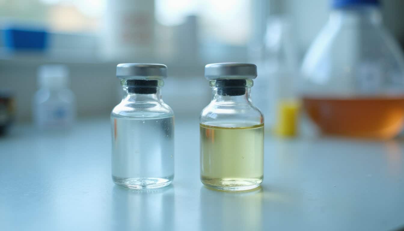
If you are looking for laboratory-grade peptides that are properly handled, securely packaged, and shipped with care, Holas has you covered. We provide research-grade peptides with third-party tested purity, reliable packaging standards, and fast shipping to support your research needs. Browse our full catalog or contact us to find the right materials for your research today. All products are for laboratory research use only and are not for human or veterinary use.
Frequently Asked Questions
What Concentration of GHK-Cu Is Biologically Active in Laboratory Skin Models?
GHK-Cu is biologically active across a broad low-dose range in laboratory skin models. In dermal fibroblast studies, 1 to 10 nM concentrations repeatedly stimulate collagen and elastin synthesis, while low micromolar concentrations drive keratinocyte proliferation and epidermal stem cell marker expression in skin-equivalent models. At 1 nM, GHK-Cu has been shown to accelerate irradiated fibroblast growth and upregulate VEGF and bFGF production, all at non-toxic concentrations across multiple in vitro systems.
Does GHK-Cu Influence Collagen Production Differently in Aged Versus Young Fibroblasts?
Yes. GHK-Cu influences collagen production in both aged and young fibroblasts, but the greater functional restoration appears in aged cells. Aged fibroblasts treated with GHK-Cu show enhanced collagen gel contraction, improved migration, and partial reversal of senescence-associated dysfunction. Young fibroblasts still respond with increased collagen synthesis, but the relative change is smaller since their baseline regenerative capacity is already higher. The evidence supports a context-dependent effect favoring compromised cellular states.
How Does Copper Binding Specifically Contribute to GHK-Cu’s Biological Activity?
Copper binding converts the GHK tripeptide into its biologically active complex by coordinating Cu(II) through the histidine residue at high affinity. This chelation enables reversible Cu(II)/Cu(I) redox cycling, which mediates electron transfer and delivers copper to metalloenzymes such as superoxide dismutase and lysyl oxidase. Without copper binding, GHK does not activate the collagen synthesis, gene-modulation, or antioxidant defense pathways documented in research.
What Does the Research Show About GHK-Cu and Route of Delivery?
In the research record, GHK-Cu‘s collagen effects are best documented for topical and in vitro exposure, with supporting data from animal wound models. Its activation of TGF-beta signaling and fibroblast stimulation is well established in laboratory models, with robust evidence for collagen I and III upregulation in cell and tissue assays. High-quality human trials specifically validating systemic or injectable delivery remain limited, so most delivery-related conclusions are extrapolated from topical, in vitro, and animal studies rather than confirmed in controlled human trials.
Are GHK-Cu’s Gene Expression Effects Reversible After Exposure Ends?
Current evidence suggests GHK-Cu‘s gene expression effects are likely reversible once exposure ends. Because GHK-Cu functions as a transient signaling peptide rather than a gene-editing agent, its transcriptional influence depends on sustained exposure. Collagen synthesis upregulation, matrix remodeling signals, and anti-inflammatory gene shifts generally require ongoing stimulation to persist. Direct human data on post-exposure reversal remain limited, but the peptide’s mechanism supports an exposure-dependent rather than permanently altered gene expression profile.