Fibroblast proliferation assays measure whether a peptide changes the growth kinetics of human dermal fibroblasts under defined conditions. You seed HDFs at a validated density, allow 24 hours for attachment, then apply peptide dilutions against untreated, blank, and vehicle-matched controls. The common colorimetric readouts, WST-1 and MTT, measure tetrazolium reduction rather than cell number, which means an absorbance change is a metabolic signal first and a proliferation claim only after confirmation.
Key Takeaways
- Seed HDFs at a consistent density, commonly 6,000 cells/cm² for peptide-response work, and allow 24 hours attachment at 37°C with 5% CO₂.
- Apply peptide dilutions after attachment with untreated, blank, and vehicle-matched controls under matched serum conditions.
- WST-1 is added at 10 µL per 100 µL medium and read at 440 nm with a reference near 600 nm. MTT reads at 570 nm after solubilization.
- Tetrazolium assays report metabolic reduction, not cell count. A peptide that alters mitochondrial activity shifts the signal without changing proliferation.
- Confirm any consequential result with EdU incorporation or direct counts before calling an absorbance change proliferation.
Why fibroblast proliferation is a useful readout
Human dermal fibroblasts are matrix-producing stromal cells and one of the most tractable primary human cell types available. They divide reliably, tolerate 96-well format, and respond measurably to growth factors, matrix cues, and adhesion conditions, which makes proliferation a convenient first functional readout for a compound with no established mechanism.
They are also the cell type where several research peptides are most often studied. GHK-Cu work centers on HDFs, usually with collagen output as the endpoint, and proliferation runs alongside that as a control on interpretation. A collagen increase means something different if cell number also rose. BPC-157 and TB-500 both carry tissue-repair claims for which fibroblast growth kinetics are a natural in vitro entry point. HUVEC tube formation for peptides can provide insights into angiogenesis and endothelial function. Understanding how these peptides influence tube formation may help establish their potential therapeutic applications.
What proliferation does not tell you is worth stating early. A change in fibroblast number is not evidence of repair, and it is not evidence of a mechanism. It establishes that the cells respond to the compound under these conditions. Anything beyond that requires migration, adhesion, collagen synthesis, or receptor-level work run separately. Collagen synthesis assay for ghk-cu can provide valuable insights into the underlying mechanisms of tissue repair. By measuring the levels of collagen produced, researchers can better understand how these compounds influence fibroblast activity.
How do you culture fibroblasts for peptide testing
Seed HDFs into 96-well plates in assay medium at a defined density and allow 24 hours at 37°C with 5% CO₂ for attachment and stabilization before any treatment. Density is a controlled variable rather than a convenience: 6,000 cells/cm² supports rapid growth for peptide-response work, while 3,000 cells/cm² suits routine expansion before plating.
Three conditions do most of the damage when they drift:
- Confluency. Do not let wells overgrow. High confluency changes receptor signaling, nutrient demand, and peptide sensitivity, so a plate that reached confluence early is not comparable to one that did not. When expanding cells beforehand, replace growth medium every other day and plate at around 60 percent confluency.
- Passage number. HDFs are primary cells with finite replicative capacity. Growth kinetics slow at higher passage, so passage is matched across all treatments in a study, not just within a plate.
- Serum. Serum contains growth factors that compete with whatever the peptide is doing. Match serum concentration across every well, and keep it low enough that baseline proliferation has room to move.
Distribute the suspension evenly, avoid edge wells drying, confirm uniform attachment microscopically before dosing, and equilibrate peptide-containing medium to culture conditions before it goes on cells.
What methods measure fibroblast proliferation
Three method classes are in common use, and they measure different things.
| Method | What it measures | Read |
|---|---|---|
| WST-1 | Tetrazolium reduction by viable cells | 440 nm, reference ~600 nm |
| MTT | Tetrazolium reduction by viable cells | 570 nm, after solubilization |
| EdU incorporation | DNA synthesis in cycling cells | Fluorescence, per-cell counting |
WST-1. Add at 10 µL per 100 µL medium, incubate until formazan develops, shake briefly to distribute, and read. The formazan is water-soluble, so no solubilization step is required and the plate can be read directly. Include blank wells containing medium plus reagent without cells.
MTT. Incubate with the dye, then solubilize the insoluble formazan crystals before reading. The extra step introduces variability, since incomplete dissolution reads as reduced signal.
EdU. Label newly synthesized DNA, fix, detect the incorporated nucleoside by fluorescence, and count cycling cells directly. Slower and more expensive than the colorimetric methods, and the only one of the three that measures division rather than inferring it.
Why a metabolic signal is not a cell count
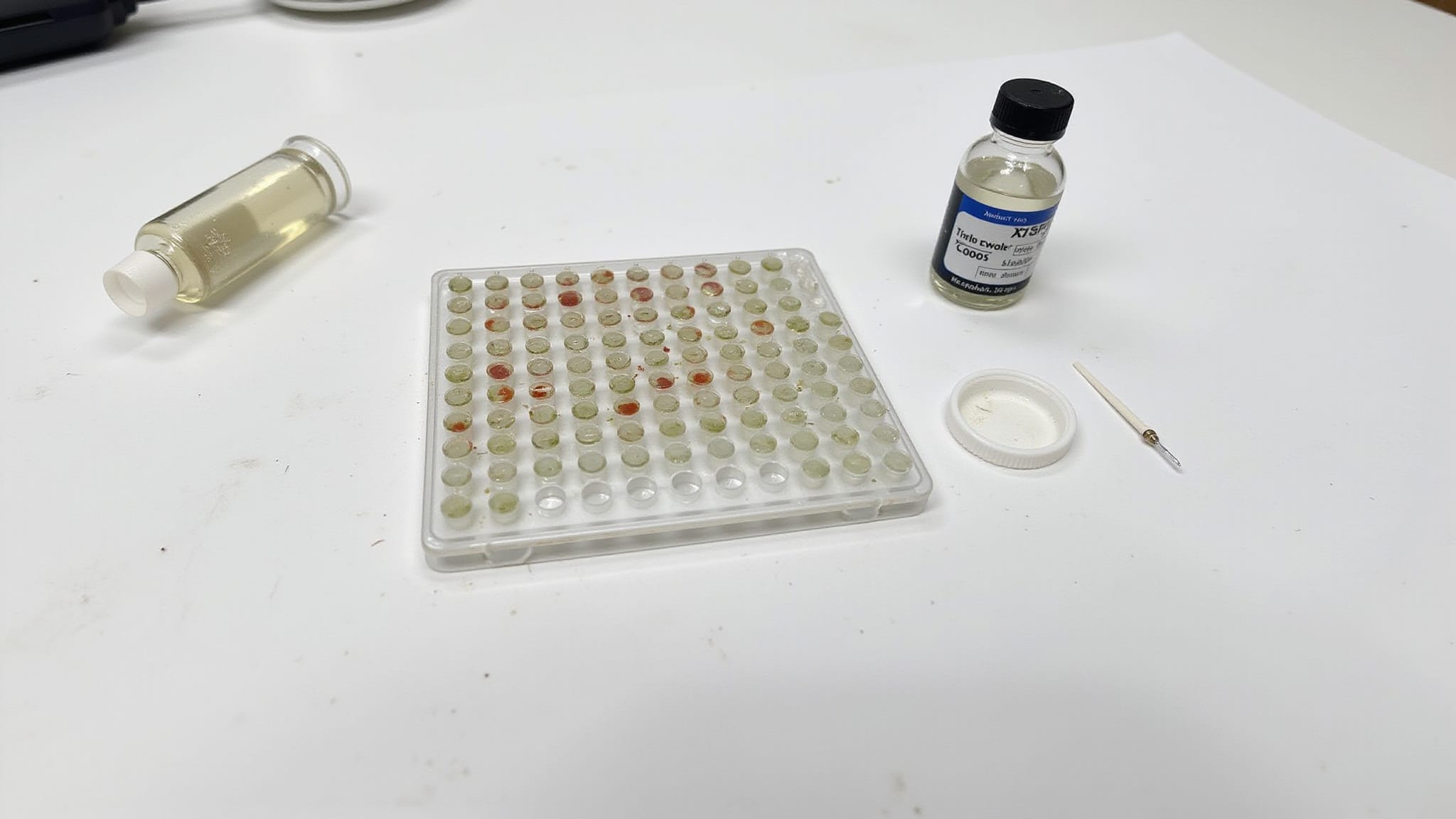
This is the assumption the assay rests on, and it is the one most often left unexamined. WST-1 and MTT quantify how much tetrazolium the well reduces. That correlates with viable cell number only when metabolic activity per cell stays constant, and a peptide is precisely the kind of intervention that can break that assumption. Mtt cell viability assay methods are often utilized in various experimental setups to gauge cellular health. Understanding the limitations of these methods is crucial as they might not account for variability in metabolic responses.
Consider what an increased absorbance reading is compatible with:
- More cells. The interpretation you want, and one of several available.
- The same cells, more active. A compound that raises mitochondrial activity or shifts cells toward oxidative metabolism increases reduction per cell without a single extra division.
- The same cells, larger. Fibroblasts that spread more on a treated surface can carry higher metabolic output per cell.
- An activation state change. Fibroblasts pushed toward a more synthetic phenotype are metabolically busier, which reads as growth.
The reverse holds too. A reduced signal is compatible with fewer cells, with metabolically quieter cells, or with cells that are alive and dividing normally while consuming less. Any of the three produces the same well.
The confound has an unhelpful property: it is dose-dependent. A peptide that alters metabolism does so more at higher concentrations, so the artifact produces a clean dose-response curve indistinguishable in shape from real proliferation. Replicate agreement, low variance, and a good curve fit offer no protection, because the artifact is as reproducible as the phenomenon.
The control is orthogonal measurement. EdU or direct counts on the key conditions, particularly whenever the compound plausibly touches metabolism, and always before an absorbance change gets reported as proliferation.
What controls and standards are essential
Four controls, each answering a distinct question.
- Blank wells. Medium plus reagent, no cells. Subtract this optical background from every reading. Reagent and medium both absorb, and the offset is not negligible.
- Untreated controls. Same density, same medium, no peptide. This sets baseline proliferation under identical incubation, CO₂, and nutrient conditions, and it is the denominator for normalization.
- Vehicle-matched controls. Same solvent concentration as the peptide wells. This matters more than it appears. Residual counterion carried in with a lyophilized peptide, or the DMSO used to dissolve it, can suppress proliferation independently of the compound, and without a matched vehicle arm that suppression is attributed to the peptide.
- Density titration. Run before the study to confirm that absorbance scales linearly with cell number across your working range. Outside the linear range, a real difference in cell number produces a compressed or absent difference in signal.
Where available, include a known mitogen as a positive control. A plate on which the positive control fails to separate from untreated is a plate that could not have detected an effect, and that is worth knowing before interpreting a negative result.
How do you analyze and interpret proliferation data
Subtract the mean blank from each reading, then express treated wells as a percentage of the untreated or vehicle control. Use replicate means with standard deviation or confidence intervals, and flag edge effects, saturation, and outliers before any statistics.
Plot dose-response curves and, where the question concerns growth rate rather than a single endpoint, time courses across 24 hours, 2 days, 5 days, and 7 days. Slope across timepoints is more informative than a single reading, because a compound that accelerates early growth and one that delays regression produce the same value at one timepoint and different curves across four.
Interpret an increased signal as proliferation only when three things hold: viability, morphology, and confluency stay consistent across wells; the reading sits inside the linear range established by your density titration; and the effect survives confirmation by EdU or counts. Absent those, the honest statement is that tetrazolium reduction increased, which is a real observation and a narrower one.
Conclusion
Fibroblast proliferation is one of the easiest assays to run and one of the easiest to over-read. The colorimetric methods that make it convenient, WST-1 and MTT, do not count cells. They measure how much tetrazolium a well reduces, and they stand in for cell number only while metabolic activity per cell holds steady. A peptide is exactly the kind of thing that can move that.
Two habits carry most of the reliability. Run a vehicle-matched control, since solvent and residual counterion suppress proliferation on their own and the suppression otherwise lands on the compound. And confirm any consequential result orthogonally, because a metabolic artifact scales with dose and produces the same clean curve a real effect would.
The claim the assay supports is narrow: fibroblasts respond to this compound, at these concentrations, under these conditions. Whether they divided more, worked harder, or changed state is a separate question, and only a separate measurement answers it.
Shop Research Peptides at Holas Today
If you are looking for research peptides that are properly handled, securely packaged, and shipped with care, Holas has you covered. We provide laboratory-grade peptides with third-party tested purity, reliable packaging standards, and fast shipping to support your research needs. Browse our full catalog or contact us to source the right peptides for your protocol.
Frequently Asked Questions
What density should HDFs be seeded at?
6,000 cells/cm² supports rapid growth for peptide-response assays, and 3,000 cells/cm² suits routine expansion before plating. Density is matched across every condition in a study, since it shifts baseline proliferation and peptide sensitivity independently of treatment.
At what wavelength is WST-1 read?
440 nm, with a reference read near 600 nm. WST-1 formazan is water-soluble and requires no solubilization step. MTT is different: it reads at 570 nm and requires crystals to be dissolved first.
Does a higher absorbance reading mean more cells?
Not necessarily. Tetrazolium assays measure metabolic reduction, which tracks cell number only if activity per cell stays constant. A compound that raises mitochondrial activity, increases cell spreading, or shifts fibroblasts toward a more synthetic phenotype increases the signal without additional division.
Why confirm with EdU or cell counts?
Because a metabolic artifact is dose-dependent and reproducible, so it produces a clean dose-response curve indistinguishable from real proliferation. Replicate agreement and a good curve fit do not separate the two. EdU measures DNA synthesis directly and counts measure cell number directly.
Why is a vehicle-matched control necessary?
Solvent and residual counterion carried in with a lyophilized peptide can suppress proliferation on their own. Without a control arm containing the same vehicle at the same concentration, that suppression is attributed to the peptide.
Why does confluency matter before dosing?
High confluency changes receptor signaling, nutrient demand, and peptide sensitivity. A plate that approached confluence before treatment is not comparable to one that did not, so cells are plated at around 60 percent confluency and wells are not allowed to overgrow.
Does increased fibroblast proliferation demonstrate a mechanism?
No. Proliferation is an integrated output of signaling, adhesion, and survival. A change establishes that the cells respond to the compound under these conditions. Attribution to a pathway requires orthogonal work such as receptor blockade, knockdown, or phosphorylation readouts.