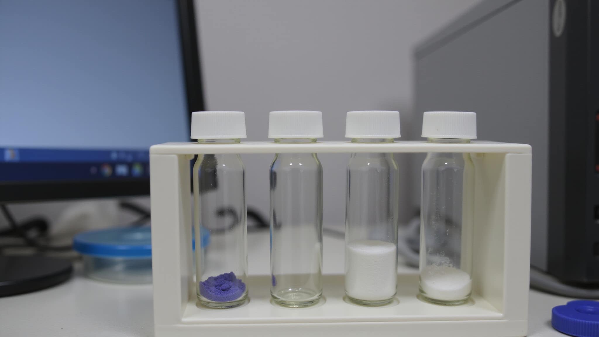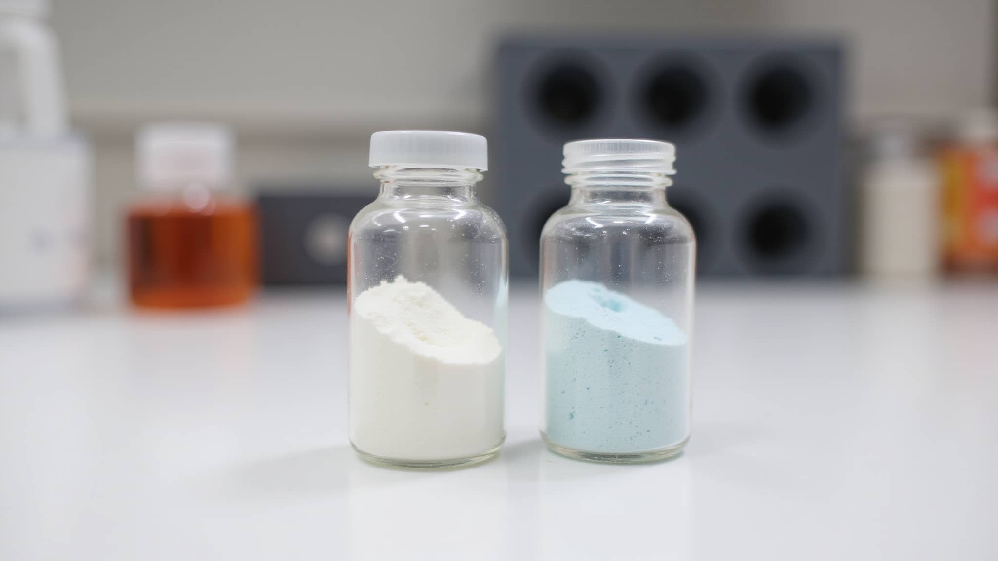
Counterion analysis confirms the actual salt form of a peptide, not just the label claim. You quantify residual anions such as trifluoroacetate, acetate, chloride, or formate, usually by ion chromatography with calibrated standards, then report results as % w/w and molar equivalents per peptide charge site. Because counterions occupy real mass in the vial and can carry their own biological activity, salt form affects mass balance, assay interpretation, and lot-to-lot comparability.
Key Takeaways
- Most research peptides arrive as TFA salts, because trifluoroacetic acid is standard in reversed-phase HPLC purification.
- Counterion content occupies measurable mass, so a milligram of TFA salt contains less peptide than a milligram of free base.
- Residual TFA is not inert in cell culture and can depress viability readings independently of the peptide.
- Ion chromatography with conductivity detection quantifies trifluoroacetate, acetate, chloride, and formate against external standards.
- Report as molar equivalents per peptide charge site alongside percent weight, so lots are compared on the same basis.
What are counterions and why analyze them
Counterions are oppositely charged ions that balance the charged sites on a peptide salt. Every lyophilized peptide with basic residues carries them, and they are not a trace contaminant. In a basic-rich sequence, counterion mass can account for a meaningful fraction of the powder in the vial.
That has an immediate practical consequence. If you weigh out 5 mg of a TFA salt and calculate a molar concentration using the peptide’s free-base molecular weight, your actual concentration is lower than your intended one, because part of what you weighed was trifluoroacetate and part was residual water. The error is systematic, it propagates through every dilution in the series, and nothing in the experiment flags it.
Counterion analysis answers three questions that purity data cannot:
- What salt form is this actually. A label stating TFA salt is a claim about the process, not a measurement of the product.
- How much peptide is in the vial. Net peptide content requires counterion content, water content, and purity together. Any one alone is insufficient.
- Are these lots comparable. Two lots at the same stated purity can carry different counterion burdens, which shifts effective concentration between them.
What is TFA and why is it in your peptide

TFA is trifluoroacetic acid, a strong, volatile acid used throughout peptide synthesis and reversed-phase HPLC purification. It appears in the finished product for a straightforward reason: it is in the mobile phase during the last purification step, and it does not entirely leave during lyophilization.
TFA serves two distinct functions, and they are worth separating because they are often collapsed into one.
Chromatographic. TFA improves peak shape across sequences by suppressing silanol interactions on the stationary phase. This sharpens resolution and improves retention reproducibility regardless of the peptide’s composition. Peptide secondary structure analysis reveals vital information about the conformation of peptides, which can significantly influence their biological activity.
Ion-pairing. TFA pairs with protonated basic side chains. Sequences rich in Arg, His, or Lys have more cationic sites and therefore carry a proportionally higher TFA burden into the lyophilized product. Sequences with few basic residues carry less.
After lyophilization, residual TFA remains as trifluoroacetate paired with those cationic sites. This is why most research peptides are supplied as TFA salts by default. It is a consequence of standard preparative workflow rather than a defect in it. Stability of lyophilized peptides is crucial for their effectiveness in various applications. When properly stored, these peptides can maintain their integrity over extended periods.
Acetate salts exist as an alternative, produced by ion exchange after purification. Acetate is generally preferred where residual TFA would confound the intended measurement, and the tradeoff is additional processing and some peptide loss.
How does residual TFA affect cell-based assays
This is the part most often missed. Residual TFA is not a passive passenger. At levels routinely present in unexchanged material, trifluoroacetate can suppress proliferation and reduce viability readings on its own.
The failure mode should look familiar to anyone who has chased a contamination artifact. A peptide is screened for cytotoxicity, viability drops in a concentration-dependent way, replicates agree, and the dose-response curve fits cleanly. Every internal consistency check passes. But serial dilution of the peptide serially dilutes the TFA alongside it, so the curve you are fitting may belong to the counterion rather than the compound. Endotoxin and research peptides can introduce additional layers of complexity when analyzing biological responses. Understanding the interaction between these components is crucial for obtaining accurate experimental results.
Several conditions make the confound harder to catch:
- It scales with the peptide. Because counterion and peptide are diluted together, the artifact produces exactly the curve shape a real effect would.
- It is invisible to purity data. A chromatogram reporting 98 percent purity says nothing about how much trifluoroacetate accompanies that 98 percent.
- It varies by sequence. A basic-rich peptide carries more TFA than a neutral one at the same stated purity, so the artifact is stronger for some compounds than others.
- It shifts between lots. Two lots from the same synthesis route can differ in residual TFA, which reads as lot-to-lot variability in biological activity.
The control is a vehicle arm containing the equivalent counterion concentration without the peptide. If viability drops in that arm, the effect is at least partly salt. Without it, a cytotoxicity result from unexchanged material carries an alternative explanation the data cannot separate.
How do you measure counterion content in peptides
Counterion content is measured by ion chromatography with conductivity detection, quantifying residual anions against calibrated external standards and verifying mass balance against peptide content and water.
- Prepare the sample. Dissolve a known peptide mass in validated diluent, record concentration precisely, and filter particulates. The mass measurement is where most error enters.
- Separate the anions. Run IC with conductivity detection to resolve trifluoroacetate, acetate, chloride, fluoride, and formate. Trifluoroacetate and acetate elute at different retention times and are distinguished by comparison to standards.
- Calibrate the response. Use external standards across the expected range. Confirm linearity, retention time reproducibility, and recovery before accepting sample values.
- Report the result. Express each ion as % w/w and as molar equivalents per peptide, since the two answer different questions.
Where IC is unavailable, validated HPLC or GC methods can substitute, though IC is the more direct approach for this analyte class.
How should counterion results be reported
There is no universal counterion cutoff, because what counts as acceptable depends on what the material is being used for. What matters more than the number is the basis it is reported on.
| Reporting basis | What it tells you | When to use it |
|---|---|---|
| Percent weight (% w/w) | How much vial mass is counterion | Calculating net peptide content |
| Molar equivalents per peptide | How saturated the charge sites are | Comparing sequences with different charge counts |
| Ion-to-peptide ratio | Stoichiometry of the salt as isolated | Assessing exchange completeness |
Percent weight alone is misleading across sequences. A peptide with six basic residues and one with two will differ substantially in % w/w TFA at identical exchange efficiency, purely because they have different numbers of sites to fill. Molar equivalents normalizes for that, which is why stoichiometric reporting is the more useful basis for comparison.
The practical rule is to pick one basis and hold it. Lots compared on different bases are not being compared.
What else does residual TFA interfere with
Cell viability is the most consequential interference, but not the only one.
FTIR amide I. The trifluoroacetate carbonyl absorbs near 1673 cm⁻¹, directly overlapping the amide I region used to assign secondary structure. Residual TFA distorts that band and can produce misassignment, which is why counterion content matters before infrared structural work.
Solution pH. Elevated TFA lowers the pH of a reconstituted preparation. In a weakly buffered system, that shift alone can alter peptide conformation and apparent behavior.
Conformational readouts. Because trifluoroacetate interacts with charged side chains, it can influence hydrogen-bonding networks and apparent helical content. A structural measurement made on unexchanged material is measuring the salt as much as the sequence.
Mass balance. Counterion mass, residual water, and purity together determine net peptide content. Reporting any one without the others gives an incomplete picture of what is in the vial.
Conclusion
Salt form is the least discussed property of a research peptide and one of the more consequential ones. It determines how much peptide is actually in the vial, it introduces a compound with its own biological activity into every well, and it does both silently, because nothing on a chromatogram or a mass spectrum reports it.
Two habits address most of the exposure. Calculate concentrations from net peptide content rather than from powder mass, which requires counterion and water data alongside purity. And when working with unexchanged material in cell-based assays, run a counterion-matched vehicle control, because a dose-response curve built on serial dilution dilutes the salt in perfect step with the peptide and will not reveal the difference on its own.
Counterion analysis converts an assumption into a measurement. You don’t just release a peptide, you release a characterized material.
Shop Research Peptides at Holas Today
When you need research peptides that are handled properly, sealed securely, and delivered with care, Holas is the trusted source your lab needs. We supply laboratory-grade peptides backed by third-party tested purity, dependable packaging standards, and fast shipping designed to keep your research moving forward. Browse our full catalog or contact us to find the right peptides for your work today.
Frequently Asked Questions
Why are most research peptides supplied as TFA salts?
Trifluoroacetic acid is standard in the mobile phase during reversed-phase HPLC purification, where it suppresses silanol interactions and sharpens peak shape. It does not fully leave during lyophilization, so it remains paired with protonated basic side chains in the finished powder. The TFA salt is a consequence of the standard purification route.
Does counterion content affect how much peptide is in the vial?
Yes. Counterion mass, residual water, and purity together determine net peptide content. Weighing out a TFA salt and calculating molarity from the free-base molecular weight overstates the actual concentration, and the error propagates through every dilution in the series.
Which peptides carry the most TFA?
Sequences rich in Arg, His, or Lys. TFA ion-pairs with protonated basic side chains, so more cationic sites means a proportionally higher trifluoroacetate burden. Sequences with few basic residues carry less at the same purity.
Can residual TFA affect cell viability results?
Yes. Trifluoroacetate can suppress proliferation at levels routinely present in unexchanged material. Because serial dilution of the peptide dilutes the counterion alongside it, the artifact produces a clean concentration-dependent curve. A counterion-matched vehicle control is what separates the two.
How is counterion content measured?
Ion chromatography with conductivity detection, quantifying trifluoroacetate, acetate, chloride, and formate against calibrated external standards. Trifluoroacetate and acetate resolve at different retention times and are identified by comparison to reference standards.
Should results be reported as percent weight or molar equivalents?
Both, because they answer different questions. Percent weight tells you how much vial mass is counterion, which matters for net peptide calculations. Molar equivalents per peptide normalizes for charge count, which is the only basis on which sequences with different numbers of basic residues can be meaningfully compared.
Why does TFA interfere with FTIR secondary structure analysis?
The trifluoroacetate carbonyl absorbs near 1673 cm⁻¹, overlapping the amide I region used to assign secondary structure. Residual TFA distorts that band and can lead to misassignment, so counterion content matters before infrared structural work.